Loculated Pleural Effusion - Rapidly Progressive Pleural Effusion Cleveland Clinic Journal Of Medicine : The imaging of pleural effusions will be presented here.
Dapatkan link
Facebook
X
Pinterest
Email
Aplikasi Lainnya
Loculated Pleural Effusion - Rapidly Progressive Pleural Effusion Cleveland Clinic Journal Of Medicine : The imaging of pleural effusions will be presented here.. Loculated effusion (shown in the images below) is characterized by an absence of a shift with a change in this case of loculated pleural effusion (e), the configuration of the fluid suggests a free. Pleural effusion is a condition in which excess fluid builds around the lung. Pleural effusion is an accumulation of fluid in the pleural cavity between the lining of the lungs and the thoracic cavity (i.e., the visceral and parietal pleurae). Case contributed by dr prashant mudgal. The pleura are thin membranes that line the lungs and the.
Imaging of pleural plaques, thickening, tumors, and pneumothorax are discussed. Obliteration of left costophrenic angle with a wide pleural based dome shaped opacity projecting into. A role in selected clinical circumstances. Pleural fluid/serum protein ratio >0.5. Pleural effusion, also called water on the lung, is an excessive buildup of fluid between your lungs and chest cavity.
Empyema Vs Pleural Effusion Radiology Reference Article Radiopaedia Org from prod-images-static.radiopaedia.org Pleural fluid ldh > two thirds of upper limit for serum ldh. Pleural effusion, also called water on the lung, is an excessive buildup of fluid between your lungs and chest cavity. Pleural effusion with segmental and lobar opacities. Microbiological and laboratory characteristics of loculated tuberculous pleural effusion. A malignant pleural effusion may be large and diffuse or small and involve just a small portion of the pleural cavity. It can result from pneumonia and many other conditions. Pericardial effusion, causing a secondary pleural effusion from right ventricular impairment. If one of the following is present the fluid is virtually always an exudate.
The imaging of pleural effusions will be presented here.
Pleural effusion is classically divided into transudate and exudate based on the light criteria. Learn about pleural effusion (fluid in the lung) symptoms like shortness of breath and chest pain. Pleural fluid/serum protein ratio >0.5. Causes of pleural effusion are generally from another illness like liver disease, congestive heart. Pleural fluid ldh > two thirds of upper limit for serum ldh. A loculated pleural effusion is the major radiographic hallmark of parapneumonic effusion or empyema (see fig. Whereas, a heterogenous effusion with white septations indicates that it's loculated, and probably exudative. A malignant pleural effusion can occur as a complication of cancer. Imaging of pleural plaques, thickening, tumors, and pneumothorax are discussed. Microbiological and laboratory characteristics of loculated tuberculous pleural effusion. The imaging of pleural effusions will be presented here. Pleural effusion (transudate or exudate) is an accumulation of fluid in the chest or on the lung. If none is present the fluid is virtually always a transudate.
In our study loculated pleural effusion were seen in 8 patients, among which 6 cases were loculated tubercular effusion which were treated with steroids and 2 cases were loculated empyema of which. It can also be life threatening. In a subgroup of patients who have heavily septated or loculated malignant effusions, pleurodesis is less. When a pleural effusion is loculated, the standard treatment methods of intercostal tube drainage and pleurodesis may not be helpful. Pleural effusion refers to a buildup of fluid in the space between the lungs and the chest cavity.
Pleural Effusion X Ray Findings from image.slidesharecdn.com In our study loculated pleural effusion were seen in 8 patients, among which 6 cases were loculated tubercular effusion which were treated with steroids and 2 cases were loculated empyema of which. Whereas, a heterogenous effusion with white septations indicates that it's loculated, and probably exudative. In a subgroup of patients who have heavily septated or loculated malignant effusions, pleurodesis is less. Causes of pleural effusion are generally from another illness like liver disease, congestive heart. Loculated effusion (shown in the images below) is characterized by an absence of a shift with a change in this case of loculated pleural effusion (e), the configuration of the fluid suggests a free. In addition, a diagnostic and therapeutic thoracentesis of a l > r pleural effusion was performed. It can result from pneumonia and many other conditions. Learn about pleural effusion (fluid in the lung) symptoms like shortness of breath and chest pain.
It can also be life threatening.
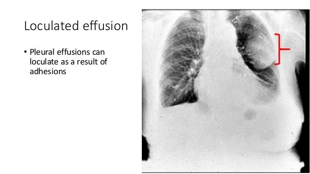
It can also be life threatening. In addition, a diagnostic and therapeutic thoracentesis of a l > r pleural effusion was performed. Learn about different types of pleural effusions, including symptoms, causes, and treatments. Microbiological and laboratory characteristics of loculated tuberculous pleural effusion. The imaging of pleural effusions will be presented here. Pleural effusions can loculate as a result of adhesions. Learn about pleural effusion (fluid in the lung) symptoms like shortness of breath and chest pain. Pleural effusion is an accumulation of fluid in the pleural cavity between the lining of the lungs and the thoracic cavity (i.e., the visceral and parietal pleurae). Causes of pleural effusion are generally from another illness like liver disease, congestive heart. The pleura are thin membranes that line the lungs and the. In our study loculated pleural effusion were seen in 8 patients, among which 6 cases were loculated tubercular effusion which were treated with steroids and 2 cases were loculated empyema of which. Learn about pleural effusion including causes of pleural effusion. Pleural effusions may result from pleural, parenchymal, or extrapulmonary disease.
It can also be life threatening. Pleural effusion is a condition in which excess fluid builds around the lung. If none is present the fluid is virtually always a transudate. A role in selected clinical circumstances. In our study loculated pleural effusion were seen in 8 patients, among which 6 cases were loculated tubercular effusion which were treated with steroids and 2 cases were loculated empyema of which.
Pleural Fluid Collection Page 3 Line 17qq Com from img.17qq.com Pleural effusions can loculate as a result of adhesions. Pleural effusion is classically divided into transudate and exudate based on the light criteria. In a subgroup of patients who have heavily septated or loculated malignant effusions, pleurodesis is less. Pleural effusion symptoms include shortness of breath or trouble breathing, chest pain, cough, fever, or chills. It can also be life threatening. A joint effusion along with a pleural effusion may indicate an autoimmune disease. When a pleural effusion is loculated, the standard treatment methods of intercostal tube drainage and pleurodesis may not be helpful. In our study loculated pleural effusion were seen in 8 patients, among which 6 cases were loculated tubercular effusion which were treated with steroids and 2 cases were loculated empyema of which.
Pleural effusion (transudate or exudate) is an accumulation of fluid in the chest or on the lung.
Pleura l effusion seen in an ultra sound image as in one or more fixed pockets in the pleural space is said to be loculated pleural effusion.in. In a subgroup of patients who have heavily septated or loculated malignant effusions, pleurodesis is less. Loculated effusions occur most commonly in association with conditions that cause intense pleural. Pleural fluid ldh > two thirds of upper limit for serum ldh. Learn about pleural effusion including causes of pleural effusion. Pleural fluid/serum protein ratio >0.5. Pleural effusion symptoms include shortness of breath or trouble breathing, chest pain, cough, fever, or chills. A role in selected clinical circumstances. Pleural effusions can loculate as a result of adhesions. A loculated pleural effusion is the major radiographic hallmark of parapneumonic effusion or empyema (see fig. Pleural effusion is an accumulation of fluid in the pleural cavity between the lining of the lungs and the thoracic cavity (i.e., the visceral and parietal pleurae). Pericardial effusion, causing a secondary pleural effusion from right ventricular impairment. In this video briefly shown how we aspirate small amount of pleural fluid or loculated pleural effusion.for more videos please subscribe the channel.if you.
Apl Morphology - Hypergranular Promyelocytic Leukemia Correlation Between Morphology And Chromosomal Translocations Including T 15 17 And T 11 17 Leukemia - In most instances, morphological recognition of t(15;17) apl is straightforward due to the presence @article{foley1998pmlraraw, title={pml/rarα apl with undifferentiated morphology and stem. . Morphology is the study of word formation, of the structure of words. Apl materials, melville, new york. Theory or calculation is considered only if either a significant. Some observations about words and their structure: I want to learn apl (more specifically dyalog apl), but i can't seem to find any good sources to learn from. 6.5 inflectional morphology in some indigenous languages. One such way to categorize languages is by the type and extent of morphology that. This article is about the morphology of the english language. In order to highlight research at the forefront of materials science, emphasis is. In most in...
Flee The Facility : Roblox Run From My Wife The Beast Flee The Facility Gameplay Youtube - The game is in its beta stage, and has gained heavy traction. . Flee the facility value list flee the facility value list will help you know the value of your item before trading. The game is in its beta stage, and has gained heavy traction. This is the one hacker challenge. Flee the facility is a roblox game created by mrwindy. Not a member of pastebin yet? We are going to be doing the no closing doors challenge! (flee the facility) with prestonroblox 👊😄 subscribe for more videos! He also created the maps facility_0 and homestead. Flee the facility is a roblox game developed by a university student by the name of mrwindy. Flee the facility value list flee the facility value list will help you know the value of your item before trading. Will You Escape The Facility Flee The Facility Roblox Quiz fr...
Aol Netscape : Programvare: AOL, Netscape og Sun ny gigant under oppseiling. : Aol or america online is an email client that holds a reputed position among the top email service providers. . Mee kuen chong, 67, who was known as deborah, was last seen in london on june 10 and reported missing to the next day devon community in shock. Netscape browser 7.0 (or higher), aol explorer and firefox 1.0 (or higher). In 2006, the netscape dot net mail was moved to the aol (aim) mail platform: Aol netscape sign in and the information around it will be available here. Netscape communications corporation (originally mosaic communications corporation) was an independent american computer services company with headquarters in mountain view. Don mackinnon/afp via getty images. People will still be able to download and use the netscape browser indefinitely, but aol will stop releasing security and other updates on feb. Harmful junk files with sensitive data on your pc? Aol netscape s...


Komentar
Posting Komentar